
Feedlot Beef Cattle Reduce Feed Intake
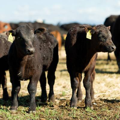
The case described is loosely based on a case submitted to UVDL. Details have been changed and added or omitted by the author.
You are a mixed-animal practitioner in rural Utah. It is a hot June day and you are called out to examine a group of feedlot beef cattle that have reduced feed intake. The producer reports that some animals are drooling and others are reluctant to move. She also says that there have been no recent changes in the feed. The producer did take the rectal temperature of several animals and she reports that it ranges from 104°F to 107°F. She also noticed some ulcerated lesions on the muzzles of two animals. Just before you end the phone call, the producer remembers that the horse she rode to look at the cattle was reluctant to take the bit when she put the bridle on and has some crusting at the corners of the mouth. She asks if you would look at the horse also when you come.
Questions
- When you get to the farm, which pens do you examine first?
- What are your main differential diagnoses?
- What is the most likely diagnosis and why? Is it a zoonotic disease?
- Once you confirm the lesions that the producer reported, what is your next step?
Case Summary
Because of the potential of a highly transmissible vesicular disease, you should examine the pens with apparently healthy animals first to avoid yourself being a fomite to transmit disease. Look at the sick animals last. Also, remember to clean and disinfect your boots and equipment between pens and change gloves. Your vet truck can also be a fomite! Good biosecurity is important.
Once you have confirmed the presence of vesicles, erosions, and oral crusting, you should call the state veterinarian (Dr. Barry Pittman) and the area veterinarian in charge (Dr. Robert DeCarolis). It is likely that they will send a foreign animal disease diagnostician (veterinarian) to conduct an investigation. Until the FADD arrives, you may ask the rancher to voluntarily quarantine the premise.
The main differential diagnoses in cattle for the scenario described above include Bovine Viral Diarrhea (BVD), Foot and Mouth Disease (FMD), Vesicular Stomatitis, Malignant Catarrhal Fever (MCF), Bluetongue, Rinderpest, Infectious Bovine Rhinotracheitis (IBR), and traumatic stomatitis. Because at least one horse is affected also, the most likely diagnosis is Vesicular Stomatitis.
Traumatic stomatitis is less likely because there have been no changes in feed (i.e. no rough forage that might injure the mouth) and because several animals are affected (traumatic stomatitis would probably be a single animal problem). Rinderpest was eradicated in 2011 so it is an unlikely diagnosis (though it should always be kept in mind in case of a possible bioterrorism event). Also, there is often diarrhea described in animals with rinderpest. In cases of MCF, there are often eye lesions in cattle and hemorrhagic enteritis, so it is a less likely diagnosis. In animals with IBR, nasal discharge, conjunctivitis, dyspnea and inflamed nares (“red nose”) are often features of the disease as well as fever and excessive salivation. Cattle with Bluetongue are often asymptomatic (it is clinically more severe in sheep) but some cattle will have fever, salivation, lacrimation, and oral vesicles/ulcers.
FMD is a foreign animal disease and it would be economically disastrous if we had an outbreak of FMD in the United States. It is, however, a disease of cloven footed animals so the presence of an affected horse makes it very unlikely to be FMD (unless the horse and cattle had encountered two different viruses). Nevertheless, all of us should be diligent in keeping an eye out for foreign animal diseases. BVD is quite common and clinical signs seen include fever, diarrhea, respiratory disease and mucosal erosions; however, it is also a disease of even-toed ungulates (which does not include horses of course!).
If you have not already figured it out, the diagnosis is Vesicular Stomatitis. Vesicular Stomatitis (VS) is caused by a virus (a rhabdovirus, the same type of virus that causes rabies). The disease occurs in horses, donkeys, mules, cattle, and swine. It is endemic in Mexico, Central America, northern South America, eastern Brazil, and limited areas of the southeastern United States. In the southwest U.S. we have periodic epidemics caused by newly introduced viruses that disappear after a couple of years. The outbreaks occur every few years and tend to end with the first frost. There are two major serotypes of the virus: New Jersey and Indiana. The lesions are very similar to FMD. Like FMD, there is high morbidity but low mortality. It is not an exotic disease, but it is reportable! Affected herds and premises are usually quarantined. The transmission of the virus is incompletely understood. Insect vectors are believed to introduce the virus into susceptible livestock and then it spreads animal to animal by direct contact. Virus is shed in vesicle fluid, saliva, and nasal secretions and also on fomites. The incubation period between infection and onset of clinical signs is 2-8 days. In some outbreaks, horses are mostly infected and in others there are more cattle affected. For example, in a large outbreak in 2005, 104 premises in Utah were quarantined and the total number of herds quarantined in the U.S. was over 580 equine facilities and over 200 bovine herds. In 2015, there were 54 infected premises in several states (Arizona, Colorado, New Mexico, Texas, and Utah). Recent changes in regulation from the USDA means that VS is delisted as a foreign animal disease in horses and quarantines of horse facilities may be lifted 14 days after the onset of clinical signs of the last affected horse (it used to be a 21-day period). However, in cattle, VS is still listed as a foreign animal disease. But remember that all cases of vesicular disease in livestock (including horses) should be reported to the state and federal veterinarians!
Vesicular stomatitis is a potentially zoonotic disease. Rarely, people exposed to the virus may develop an acute flu-like illness and occasionally vesicles may be found on the mouth, lips, or hands. Protective clothing and gloves should be worn when handling infected animals and a biosafety cabinet should be used in the lab.