
Unexpected Lamb Deaths
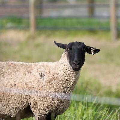
The case described is loosely based on a case submitted to UVDL. Details have been changed and added or omitted by the author.
A sheep rancher with over 30 years of experience raising lambs lost several lambs in the spring and early summer of 2018. She had never seen clinical signs and death loss like this in all her years of farming. Clinical signs were only seen in the orphan or “bummer” lambs that were fed lamb milk replacer rather than ewe’s milk. The lambs nursing ewes and adult animals on the farm were not affected. Clinical signs seen included lameness, weakness, and unwillingness to move. When the rancher examined the lambs, the lambs were in a great deal of pain and she could feel fractures of the legs.
The lambs continued to have a good appetite and were alert. With nursing care, some of the lambs recovered but, unfortunately, some were severely affected and were submitted to the UVDL, Spanish Fork laboratory for necropsy. Gross lesions seen in two 3-month-old lambs necropsied included fractures of the legs and ribs and accompanying soft tissue hemorrhage. Joints were normal and there was no evidence of disease of the major abdominal and thoracic organs. The affected lambs were housed indoors, separately from the rest of the flock, so it was unlikely that trauma (such as headbutting by ewes) was the cause of the fractures. All lambs (orphan and nursing ewes) had access to the same lamb starter grain product and the orphan lambs occasionally received Powerade mixed in the milk replacer.
Case Summary
Pathologic fractures secondary to metabolic bone disease (such as rickets) were strongly suspected as the cause of clinical signs and diseases. However, rickets is a complicated disease and documenting the cause is sometimes far from simple! Histologic lesions of the bone in the two lambs included failure of endochondral ossification and diffuse osteopenia. These lesions are consistent with metabolic bone disease. Metabolic bone diseases seldom result from a deficiency of a single dietary nutrient: often there is a deficiency of more than one nutrient or an imbalance in the calcium to phosphorus ratio. Enteric disease can affect bone development due to malabsorption of nutrients and reduced availability of the active form of vitamin D can also cause defective bone development. The most common causes of metabolic bone disease in domestic animals are dietary deficiencies of either phosphorus or vitamin D. Vitamin D needs to undergo hydroxylation, first in the liver and then in the kidney to form the active hormone 1,25-dihydroxyvitamin D which acts with parathyroid hormone to maintain plasma calcium concentrations. The active form of vitamin D enhances absorption of calcium and phosphorus from the small intestine, stimulates release of calcium and phosphorus from bone, and enhances phosphate reabsorption from the kidney. A dietary calcium: phosphorus ration of 1:1 to 2:1 is ideal. If the ratio is wider, increased vitamin D is needed for good calcium and phosphorus retention and bone mineralization. However, if the ratio imbalance is severe, increased vitamin D may not be effective at preventing lesions.
In order to determine the cause of the metabolic bone disease in these lambs, further diagnostic testing was needed. Vitamin D deficiency due to lack of sunlight was suspected, but initially testing was focused on magnesium, calcium and phosphorus concentrations in the lambs. Serum calcium concentrations were within the normal range (10.4 mg/dL; normal range 8.6 – 12.0). Serum phosphorus was elevated (12.7 mg/dL; normal range 4.5 – 8.2). However, the serum was severely hemolyzed due to postmortem collection and it is likely that the phosphorus was elevated due to hemolysis of the sample. Therefore, bone ash analysis was performed on a rib from an affected lamb. Bone magnesium was 0.5% (normal range 0.5 to 0.8%); bone calcium was 18.5% (normal range 15 to 21% on a fat free, dry weight basis); bone phosphorus was 10.4% (normal range 9 to 11% on a fat-free, dry weight basis). However, bone phosphorus may not be an accurate indicator of dietary intake in sheep. The lamb milk replacer levels of calcium and phosphorus were also analyzed: calcium at 0.73% was very close to the minimum guaranteed analysis on the label (0.75%); the phosphorus level of 0.52% was lower than the minimum guaranteed level on the label (0.70%). Some settling of minerals may have occurred during storage (the milk replacer was initially purchased in 2017 and was stored in the barn all throughout the heat of summer). Even with reduced phosphorus in the milk replacer the calcium: phosphorus ratio was within the optimal range.
With adequate direct exposure to sunlight, ruminants do not necessarily need dietary vitamin D. Vitamin D2 (ergocalciferol) is derived from plants and vitamin D3 is produced from the provitamin 7-dehydrocholesterol which is made in the body and present in the skin, intestinal wall, and other tissues. Vitamin D3 can be derived from the diet (from cod liver oil, for example) but most is formed in the skin. Vitamin D precursors must undergo irradiation(exposure to sunlight) to be activated to D2 and D3. Vitamin D3 tends to have higher potency than vitamin D2 in birds and primates, but the potencies are similar in ruminants. Vitamin D content in the lamb liver and milk replacer were determined at an outside laboratory. The lamb liver vitamin D3 was 18.9 ng/g and the lamb milk replacer vitamin D3 was 558.2 ng/g. Both of these values are within normal limits. However, it is not clear if these values represent the active form of vitamin D3 (1,25-dehydroxyvitamin D3) or the less active non-hydroxylated form. An antemortem serum sample would be needed to determine if the active form of vitamin D3 was present in the samples.
Given the clinical history and findings of diagnostic tests, metabolic bone disease was considered the most likely diagnosis and the cause was lack of the active form of vitamin D secondary to a lack of exposure to sunlight.
The following final recommendations were made to the sheep producer:
- Allow orphan lambs access to sunlight for at least a couple of hours daily.
- Purchase new milk replacer every year and store it in a cool place.
- Administer injectable vitamin D to affected lambs.
Six-month follow-up: the producer reports that all other affected lambs recovered, and you would not be able to tell that they had ever had bone problems. A happy ending.
Some interesting side notes about rickets and osteomalacia (the form in adult animals) in sheep: thick-coated sheep and those with darker wool require longer periods in the sun for adequate previtamin D3 formation. Also, animals in higher latitudes in winter may not have enough ultraviolet B light exposure for conversion of provitamin 7-dehydrocholesterol to previtamin D3. Conversely, at high altitude with intense sun exposure, previtamin D3 is converted to biologically inert compounds that are sloughed off with skin cells during normal skin turnover! This could also result in vitamin D deficiency. In addition, animals grazed on immature pasture may be vitamin D deficient due to the anti-vitamin D activity of carotenes in lush pasture and green cereal crops.
Acknowledgements
Dr. Jeffery Hall (veterinary toxicologist), Dr. Arnaud VanWettere (veterinary pathologist), Dr. Jackie Kurz (veterinary pathologist) and Dr. Paul Dawson (ruminant nutritionist) for helping me with this case.
References
- Jubb, Kennedy, and Palmer’s Pathology of Domestic animals, 5th edition, 2007, M. Grant Maxie, editor. Chapter 1, Diseases of Bone.
- Belonje PC, Van Der Berg A. Failure of bone phosphorus levels to indicate dietary intake of phosphorus by sheep. Onderstepoort J Vet Res; 50: 1-2, 1983.
- Dittmer KE, Thompson KG. Vitamin D metabolism and rickets in domestic animals: a review. Vet Path; 48: 389-407, 2011.
Van Saun RJ. Vitamin D-responsive rickets in neonatal lambs. Can Vet J; 45: 841-844, 2004.